
 |
 |
 |
 |
 |
 |
 |
 |
 |
 |
 |
 |
 |
Tree
Fern Stem
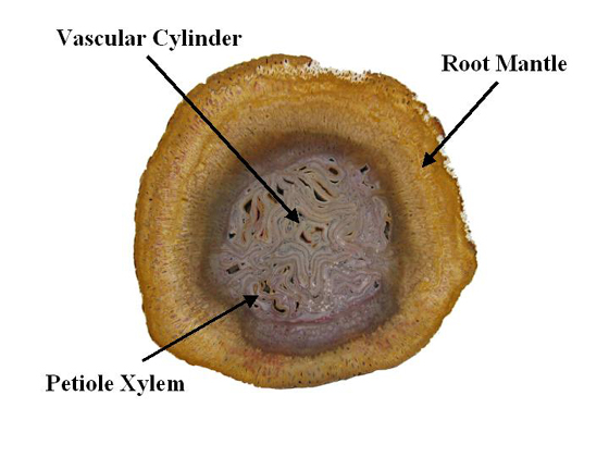 |
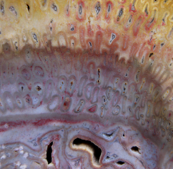
| The central vascular cylinder of the tree fern is composed of a complex arrangement of banded-shaped xylem strands. At the periphery of the central vascular cylinder c-shaped vascular strands of petioles can be seen. Surrounding the vascular cylinder is a mantle of roots. The thickness of the root mantle decreases as one moves up the trunk of the tree. The specimen above is a permian aged tree fern (Psaronius brasiliensis) from Brazil. See close-ups below. |
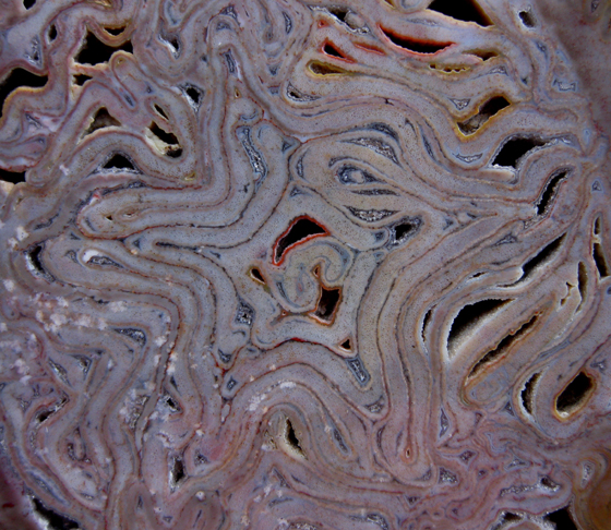 |
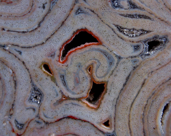
| The xylem strands in this Psaronius specimen exhibit great color and detail. The sinuous strands take the form of a pleasing star-shape. As one increases the magnification quartz crystals can be seen lining the voids. Visit the Brazil Gallery (slide 9) in the Permian section of our website to see the tracheids at 30x and 40x. |
 |
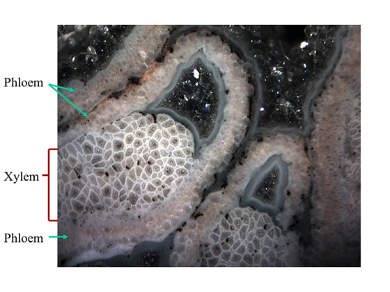
| The close-up below zooms in on a xylem strand in the photo above and was taken at 40x using a Swift M29TZ Zoom Stereo Trinocular Microscope and a Moticam 2500. The microscopes upper halogen light was used for illumination. The image was cropped and resized using Adobe Photoshop Elements 2.0. Phloem tissue can be found on both sides of the xylem tissue. |
 |
| A closer look at the transition zone between the vascular cylinder and the root mantle can be seen below. |
 |
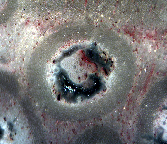
| The close-up below shows an adventitious root in the mantle of the Psaronius specimen above. Xylem cells form a star-shape at the center of the rootlet. Phloem occupies spaces between the points of the xylem star. Cortex tissue made of parenchyma cells and air spaces surround the xylem and phloem. The phloem and cortex tissue in this rootlet have been replaced by agate. A massive sclerenchyma sheath surrounds the cortex zone. Proliferating epidermis from the root and cortical tissues appear to "flow" around the individual roots which they interconnect. |
 |